 1 | 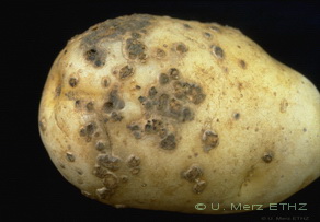 2 | 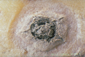 3 | 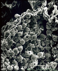 4 | 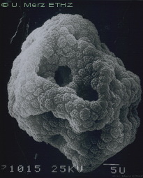 5 | 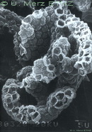 6 |
 7 | 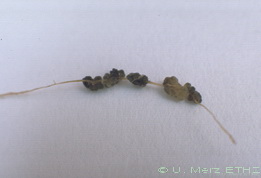 8 | 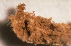 9 | 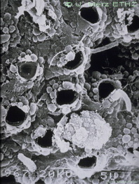 10 | 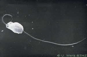 11 | 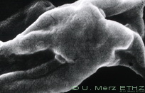 12 |
 13 | 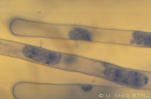 14 | 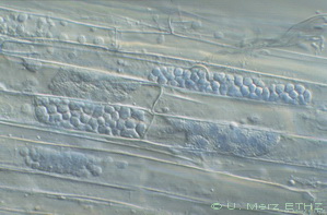 15 | 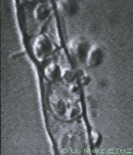 16 | 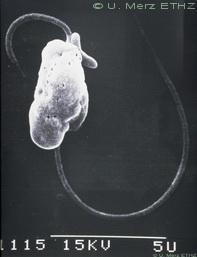 17 | 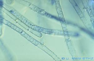 18 |
A series of DIGITAL photographs and micrographs (stereo/light/electron microscope) or individual ones can be ordered.
The complete collection consists of total 18 pictures. It will help you to make your lectures more interesting and comprehensive.
Or to brush-up your talks.
Numbered thumbnails are shown below.
1 | 2 | 3 | 4 | 5 | 6 |
7 | 8 | 9 | 10 | 11 | 12 |
13 | 14 | 15 | 16 | 17 | 18 |
Legends:
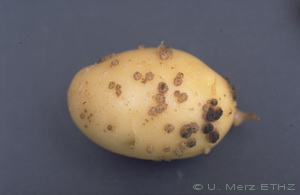
1: Young tuber with lesions in the early "cauliflower" stage
2: Tuber with typical powdery scab symptoms
3: A single lesion showing the powdery mass of resting spores (spore balls)
inside and the remnants of the broken epidermis at the edge of the lesion
4: EM-micrograph of spore balls in a lesion
5: A spore ball (sporosorus) consisting of numerous single spores
6: A broken spore ball showing the hollows and channels inside
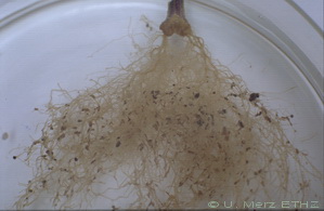
7: A root system of a potato plant (stem cutting) with root galls
8: A closer look to a piece of potato root with galls
9: A broken gall with the powdery mass of spore balls
10: A part of a resting spore ball showing single spores with an exit pores
11: A heterocont biflagellated primary zoospore
12: The two flagella are laterally inserted at an angle of about 180 degree
to each other and show a bulging basal ring at the postitions of insertion
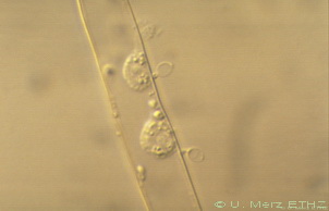
13: Tomato root hair with an encysted zoospore and the first postinfection
stage, a one-nucleus plasmodium
14: Root hairs with stained young plasmodia
15: Tomato root epidermal cells with zoosporangia of different development
stages
16: A video-captured image showing a mature zoosporangium with emerging secondary
zoospores
17: A secondary zoospore. Primary and secondary zoospores are similar in
morphology
18: Tomato plant root hairs with empty zoosporangia
Ordering informations
A complete DIGITAL picture set on a CD will include:
The price is (shipping with delivery in 2 to 3 weeks included):
Back to SPONGOSPORA Competence Center / The Genus Spongospora / Members of the Genus / References / Places and People
Copyright U. Merz, spongospora.net, CH-8408 Winterthur
Last update: November 2022